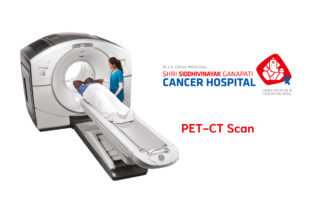
Nuclear medicine is branch of medicine which uses various radio-isotopes in diagnosis & treatment of various diseases. It is one of the most non-invasive & painless diagnostic tool to understand physiology & pathophysiology of various disease processes in the body. The contribution of nuclear medicine to overall advancement of medical sciences is immense. This is one speciality; which has literally grown from scientific laboratory to clinical bedside medicine. It has immense potential for growth as we search for answers to many questions at molecular level today.
Dr. Alok Pawaskar is working as full time consultant and head, Nuclear Medicine and PET-CT department at Shri Siddhivinayak Ganapati Cancer Hospital, Miraj. Before joining here he was consultant at HCG Manavata Cancer Hospital, Nashik for 4 years, MIOT International Hospital, Chennai for 1 year & Apollo Hospitals, Chennai for 3 yrs. He completed his graduation from V.M. Government Medical College, Solapur. He did his Diploma in Radiation Medicine (DRM) from Radiation Medicine Centre (RMC), B.A.R.C., Mumbai & DNB (Nuclear Medicine) from P. D. Hinduja National Hospitals, Mumbai. He was working at Sanjay Gandhi Post Graduate Institute of Medical Sciences (SGPGIMS), lucknow & Radiation Medicine Centre, Mumbai before moving to Chennai.
Dr. Alok Pawaskar has undergone training in PET-CT at Singapore General Hospital, Singapore. He has presented & published various papers in journals of national & International repute.
How does Nuclear Medicine works?
Most of the scans & therapies in nuclear medicine are based on tracer technique. We use a bio-molecule which is involved in particular physiological process & then tag this with radio-isotope in very small amount. Wherever this bio-molecule goes, the isotope follows it; emitting gamma rays all along. These gamma rays are then picked up by the detectors of gamma camera or PET-CT machines. The distribution of those gamma rays is then converted into an image of tracer distribution in the body; which is interpreted by nuclear medicine physician.
For therapy; similar ‘tracer’ is used which targets specific tumour cells either by uptake mechanism or by attachment to specific receptors on the tumour. This tracer carries the isotopes emitting beta particles or alpha particles which in effect specifically kill the tumour cells without much effect on normal cells.
What is so special about nuclear scans that cannot be seen by CT MRI scan?
X-ray, USG, CT scan & MRI are all anatomical imaging modalities and for that matter they do their job well. However, clinician may be interested in something more than mere size, shape & location of the organs in body. He may be able to treat in better way, if something tells him about the function of these organs ‘live’ & in quantitative manner.
Let us take an example of positron emission tomography or PET scan. FDG is a tracer which behaves similar to glucose in the body. & hence tells about glucose utilization by body organs. Now, most of the tumours need lot of glucose to grow. Hence, they stand out as areas of increased glucose consumption i.e. FDG uptake on FDG PET Scan. Further, this glucose / FDG use can be quantified, telling us which tumour is more aggressive. Routinely, on a CT scan or MRI done after treatment, sizeable amount of mass is still seen. However, CT scan or MRI cannot tell if that is a dead tissue or some tumour is still left in these masses. That is where FDG PET scan scores. It tells you about glucose used by tumour cells which are viable and simply does not show FDG uptake in dead cell masses.
This typical advantage of detecting function & functional disturbances is the strength of nuclear medicine scans. It is used to analyse & quantify function of almost every organ in the body like brain, lung, liver, kidney, bones & heart.
But you use radiation in your scans; isn’t it harmful?
Nuclear medicine has been there even before CT scanners came out in the market. So large data of evidence is available which does not show any side effects of the radiation associated with isotopes used within permissible limits for various scans & therapies in Nuclear Medicine. In fact, the amount of tracer used is typically in picomol concentrations; which is not even detected by the body to react. So, the pharmaceutical side effects one sees with contrast used in CT scan / MRI are almost nonexistent in nuclear medicine.
Of course, the precautions about right patient, right dose, right rate of administration of the right tracer apply like they do for any other medicine. And because radiation is involved, most of the Nuclear medicine procedures are not carried out in pregnant ladies. Ladies who are lactating need some precautions before they undergo the scans. Please inform your doctor if you suspect that you might be pregnant or are pregnant before undergoing any nuclear scan / therapy procedure.
What are the newer procedures done in nuclear medicine?
There is a lot of excitement about new molecules being available for PET scans. Two prominent examples are: Ga-68 DOTATATE scan for neuro endocrine tumours & Ga -68 PSMA for prostate cancer. As it stands today, for these two cancers there is no other scan as sensitive and as specific as these Ga-68 labelled tracer scans are.
Iodine-131 or I-131 labelled mIBG have been there for therapy for thyroid cancers & neuroendocrine tumours respectively for many years. Now Lu-177 labelled DOTATATE for neuroendocrine tumour & PSMA for prostate cancer are showing exciting results when every other chemotherapy / targeted therapy has failed.
Y-90 labelled microspheres are used in treatment of hepato-cellular carcinoma by directly injecting them into hepatic artery. P-32, Sm-153 have been used in bone pain palliation. Hand held gamma probe is being used for sentinel node surgeries intra operatively; so as to avoid significant morbidity in carcinoma breast or malignant melanoma patients.
The field of nuclear medicine is expanding rapidly with addition of newer tracers & techniques. With new detector materials & software, amount of isotope used for these scans is reducing drastically. It is the modality to look up to as we are entering an era of personalized medicine with its quantitative capabilities at molecular level. 

